PLOS ONE · 2026 · peer-reviewed
Agreement with a trained observer, across 1,058 dogs and 80 breeds
The measurement Workup runs is not something we are asking you to take on faith. It was validated in a retrospective method-comparison study with the Texas A&M College of Veterinary Medicine, and published where anyone can check it.
doi:10.1371/journal.pone.0337679
What it found
Mean bias and the limits of agreement against the human observer, in vertebrae. Only the published aggregates are drawn — the per-animal data is not ours to redraw, and inventing plausible points would be fabricating clinical evidence.
Limits of agreement -0.85 to 0.85 v
Difference from the trained observer (Workup − observer), in vertebrae. The center line is perfect agreement.
VHS: Pearson r 0.917, mean bias 0.002 vertebrae, limits of agreement -0.85 to 0.85 vertebrae against a trained human observer.
Limits of agreement -0.44 to 0.46 v
Difference from the trained observer (Workup − observer), in vertebrae. The center line is perfect agreement.
VLAS: Pearson r 0.873, mean bias 0.007 vertebrae, limits of agreement -0.44 to 0.46 vertebrae against a trained human observer.
- VHS · Pearson r
- 0.917
- Mean bias +0.002 v, limits of agreement -0.85 to 0.85 v.
- VLAS · Pearson r
- 0.873
- Mean bias +0.007 v, limits of agreement -0.44 to 0.46 v.
How a capture works
No export from the viewer, no manual measurement, no calipers. Workup keeps collecting until three frames are good enough, then reports how far they agreed.
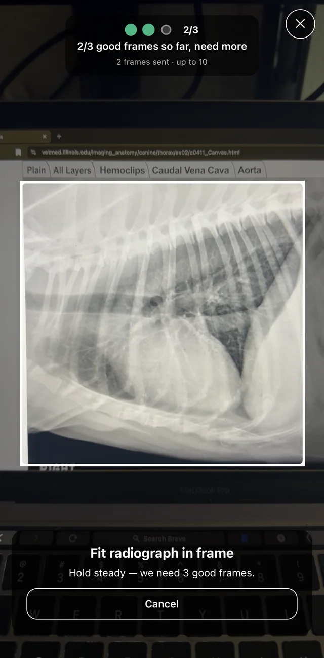
Step 1 · Capture
Point the camera at the radiograph on any monitor. Workup keeps collecting until three frames are good enough — no manual focus, no export from the viewer.
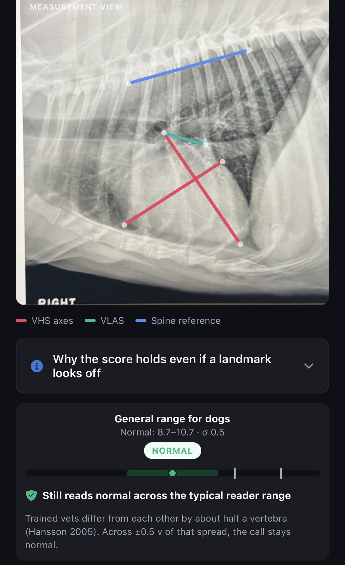
Step 2 · Measure
VHS axes, VLAS, and the spine reference drawn on your image, with the landmarks editable. The range card shows whether the call survives normal reader-to-reader variation.
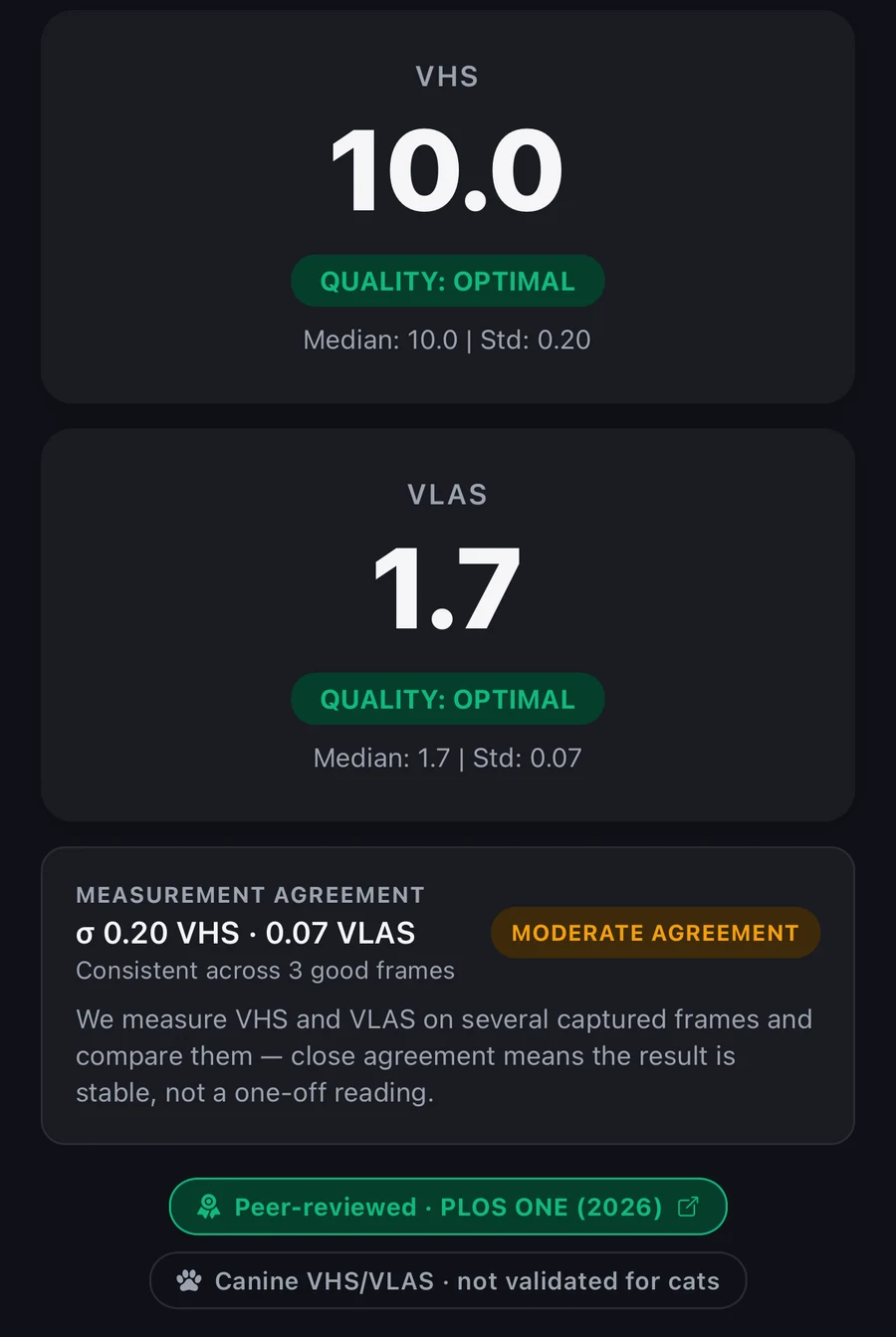
Step 3 · Read the number
VHS 10.0 and VLAS 1.7, each at optimal quality — and, reported separately, how far the three frames agreed with each other: σ 0.20 and 0.07, flagged here as moderate. Image quality and frame agreement are two different readings, so you can see when the frames disagree even though each one was clean. The canine-only limit stays on the label.
And what it does not show
A published result is only useful if its edges are stated as plainly as its headline. These are the study’s own limits.
- Design
- Retrospective, single-center method-comparison study.
- Compared to
- One trained human observer. Not a panel, not board-certified radiologists, and not echocardiography — the paper names comparison against echocardiographic measures of cardiac size as future work.
- Sample
- 1,058 client-owned dogs across 80 breeds, varied heart sizes and thoracic conformations.
- Image scope
- Good-quality right lateral radiographs, dogs only. The published validation does not extend to other views, other species, or poor-quality captures.
- Conclusion
- Differences from the human observer were of clinically insignificant magnitude, on good-quality right lateral radiographs, across dogs of all sizes.
The measurement informs your staging. It does not make it
Workup is decision support for licensed veterinary professionals and is not FDA- or USDA-cleared as a medical device. VHS/VLAS is validated for canine, good-quality right lateral radiographs only, and it runs in the cloud, so it needs a connection.
Start your 30-day free trial